Most hair-loss workups run the same generic panel for everyone. The right blood test for hair loss depends on what you are actually seeing, sudden diffuse shedding, gradual patterned thinning, postpartum loss, patchy spots, or thinning alongside other symptoms. This guide gives you the five core tests every workup should include, then routes you to the scenario-specific add-ons and the male-versus-female differences, so you walk into your appointment knowing exactly which tests to request and what each result reveals.

Five blood tests belong in almost every hair-loss workup, ferritin, thyroid-stimulating hormone, twenty-five-hydroxyvitamin D, a complete blood count, and DHT or free testosterone. The right blood test for hair loss depends on the pattern of your shedding, not on a single one-size-fits-all list, and most pages get that part wrong. This guide gives you a checklist you can hand to your GP or dermatologist, and it gives a partner researching on someone’s behalf the exact panel to request. By the end you will know not just what a blood test for hair loss measures, but which version of the panel fits what you are actually seeing in the mirror.
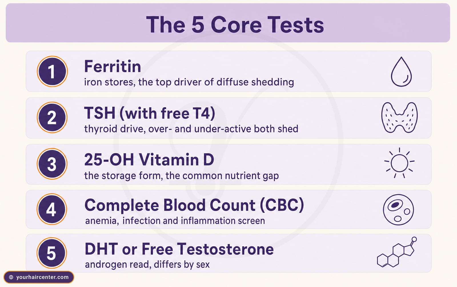
The 5 core tests in any blood test for hair loss panel
- Ferritin is the most sensitive marker of your iron stores, and low ferritin is one of the most under-diagnosed drivers of diffuse shedding, especially in menstruating women.
- Thyroid-stimulating hormone (TSH) with a free T4 reflex, because both underactive and overactive thyroid states shed hair, and TSH measured alone can miss cases that run with a low free T4.
- Twenty-five-hydroxyvitamin D (25-OH vitamin D), the storage form of the vitamin; the active form (1,25-OH) is the wrong test to order for a hair workup.
- Complete blood count (CBC), which screens for the anemia, infection, and inflammation that can push follicles prematurely into the resting (telogen) phase.
- DHT or free testosterone, read differently by sex; in men an indirect signal of androgen-driven miniaturization, in women an early flag for PCOS or other androgen pathology.
Those five anchor any sensible hair loss blood test panel. Beyond them sits an optional add-on tier, ANA, CRP, ESR, anti-TPO antibodies, zinc, vitamin B12, prolactin, the free-androgen index, and reverse T3. The add-ons are scenario-driven rather than routine, which is exactly what the next five sections walk through. At Your Hair Center, these same five core tests plus the scenario-appropriate add-ons are drawn during the supporting-labs step of the hair and scalp analysis and read alongside the trichoscopy images, so the visible state of the follicles is interpreted together with the labs rather than in isolation. That pairing is what turns a generic blood test for hair loss into a result matched to your actual pattern.
The add-on tier, what each extra marker is for
The add-ons are not a longer version of the core panel. Each one answers a specific clinical question, and a good clinician orders it only when the picture calls for it. Here is the qualitative read on each marker in this blood test for hair loss tier, so you understand why it would be added rather than assuming more tests is always better.
- Antinuclear antibody (ANA) screens for systemic autoimmune disease such as lupus, worth adding when the shedding sits alongside joint, skin, or fatigue symptoms.
- C-reactive protein (CRP) flags active inflammation anywhere in the body, useful when an immune flare is suspected behind the shed.
- Erythrocyte sedimentation rate (ESR) is a second, slower inflammation marker that complements CRP and helps gauge whether a flare is settling or persisting.
- Anti-thyroid peroxidase (anti-TPO) antibodies detect autoimmune thyroid disease before the standard TSH has drifted, which is why it earns a place in postpartum and patchy-loss workups.
- Zinc is investigated when diet, gut absorption, or restrictive eating raises the possibility of a trace-mineral gap, though routine testing is debated.
- Vitamin B12 matters most in vegans, older adults, and anyone with absorption issues, since a quiet deficiency can drive diffuse shedding.
- Prolactin is added when an elevated level is plausible, because high prolactin disrupts the hormonal balance that keeps follicles in the growth phase.
- Free-androgen index refines the androgen read in women by accounting for how much testosterone is actually unbound and biologically active.
- Reverse T3 captures a conversion-stress thyroid picture that a standard TSH can miss, relevant when symptoms persist despite normal-looking thyroid bloods.
Which of these get added is exactly where a blood test for hair loss stops being a fixed list and becomes a decision the clinical picture drives. The five scenarios below show how that decision plays out in practice.
Scenario 1, sudden diffuse shedding, the TE workup
The picture here is sudden. You notice a jump in shedding roughly two to four months after a trigger, childbirth, a major surgery, a high fever, a crash diet, a new medication, or severe psychological stress. The hair comes out in handfuls in the shower and on the pillow, but it thins evenly rather than in a defined pattern. This is the classic telogen effluvium presentation, and the blood test for hair loss panel here is shaped by the trigger behind it.
The tests that matter, with a one-line clinical read on each.
- Ferritin, the iron-store marker most closely tied to diffuse shedding. In the published telogen-effluvium series, average ferritin sat near 24 ng/mL in shedding patients versus about 46 ng/mL in unaffected controls, and a value below roughly 25 ng/mL marked the highest risk, so a low result can drive shedding even when it still falls inside the lab’s normal range.[1]
- TSH with a reflex free T4, because a normal-looking TSH can still sit alongside a low free T4, the pattern that gets missed when TSH is measured on its own.[2]
- Twenty-five-hydroxyvitamin D, read as deficient under 20 ng/mL, insufficient at 20 to 30, and sufficient at 30 or above.[3]
- CBC, to rule out occult anemia and the inflammation that drives follicles into telogen.
Be realistic about timing. The shedding lags the trigger by roughly twelve to sixteen weeks, and once you correct the underlying lab, regrowth can take up to six months to become visible.[4] Timing this blood test for hair loss to land after the shed has started gives the labs their best chance of catching the trigger. For the deep dive on the most-asked single marker, see vitamin D deficiency and hair loss, and for the differential against patterned loss read telogen effluvium versus androgenetic alopecia. When the workup confirms reversible shedding, PRP for hair recovery is one in-clinic option to support the regrowth phase.
Scenario 2, gradual patterned thinning, the AGA workup
This picture is slow. Men see recession at the temples and thinning at the crown; women see a widening part. The timeline is years rather than months, the hair on the pillow is not dramatically heavier, but the part line and temple shape look clearly different in photos a year apart. This is androgenetic alopecia, and here the logic of the blood test for hair loss flips. Blood tests do not diagnose AGA. The diagnosis is clinical and trichoscopic, and the bloods are ordered to rule out mimics and to map the androgen environment that will guide medical therapy.
The panel skews by sex.
- For men, total testosterone, free testosterone, sex hormone-binding globulin (SHBG), and DHT where the lab offers it. This answers the common question of which blood test required for hair loss male patients should request.
- For women, total and free testosterone, SHBG, the free-androgen index, DHEA-S, prolactin, and 17-hydroxyprogesterone if a PCOS or late-onset CAH picture is in play.
- For both sexes, ferritin and 25-OH vitamin D, because a coexisting deficiency makes a patterned-loss presentation look worse than the genetics alone would explain.
The clinical sequence is explicit. The doctor confirms AGA on trichoscopy, looking for miniaturization and hair-shaft caliber variability affecting more than twenty percent of hairs (anisotrichosis), a recognized major trichoscopic criterion for androgenetic alopecia, then uses the androgen panel to plan medical therapy and to flag any endocrine pathology that needs its own workup.[5] So in patterned loss the blood test for hair loss supports the diagnosis rather than making it. At Your Hair Center the hair and scalp analysis pairs that trichoscopy with the bloods, and when medical therapy plateaus the men’s hair transplant and women’s hair transplant pathways carry the sex-specific surgical options.
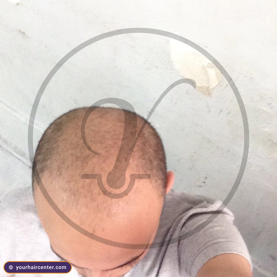
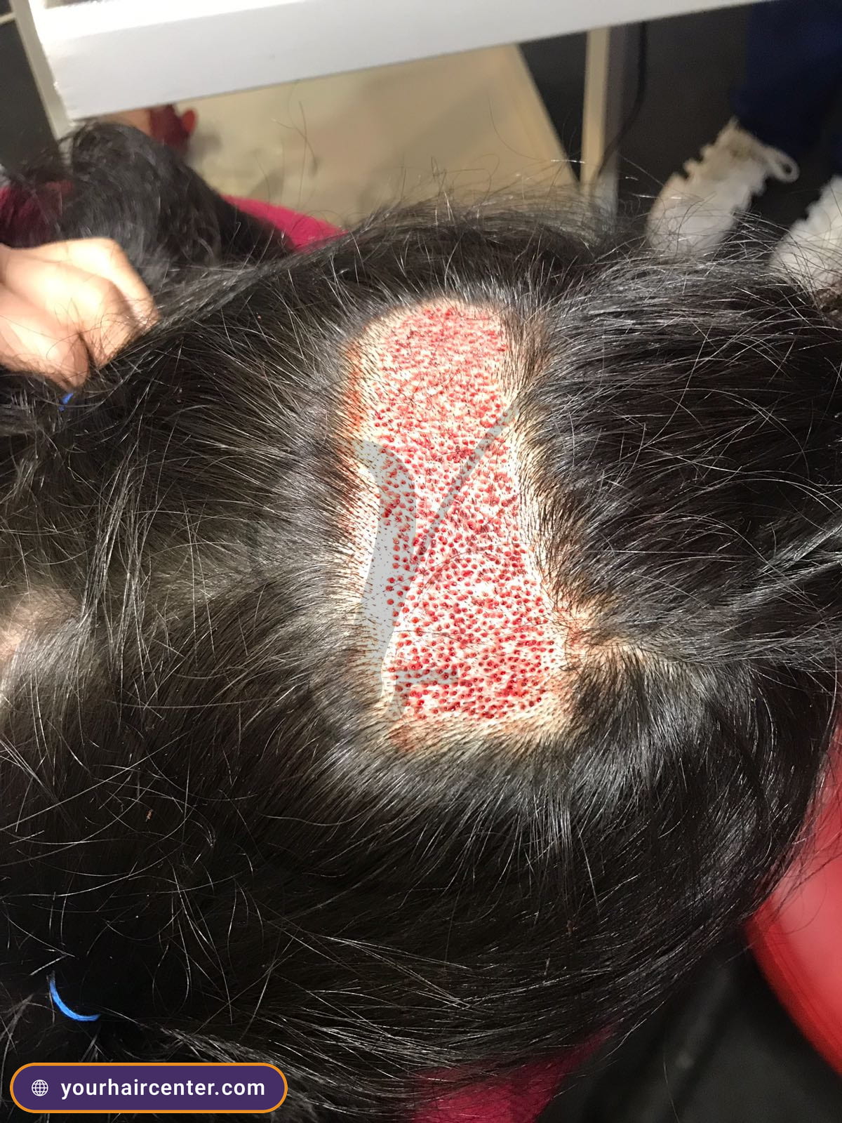
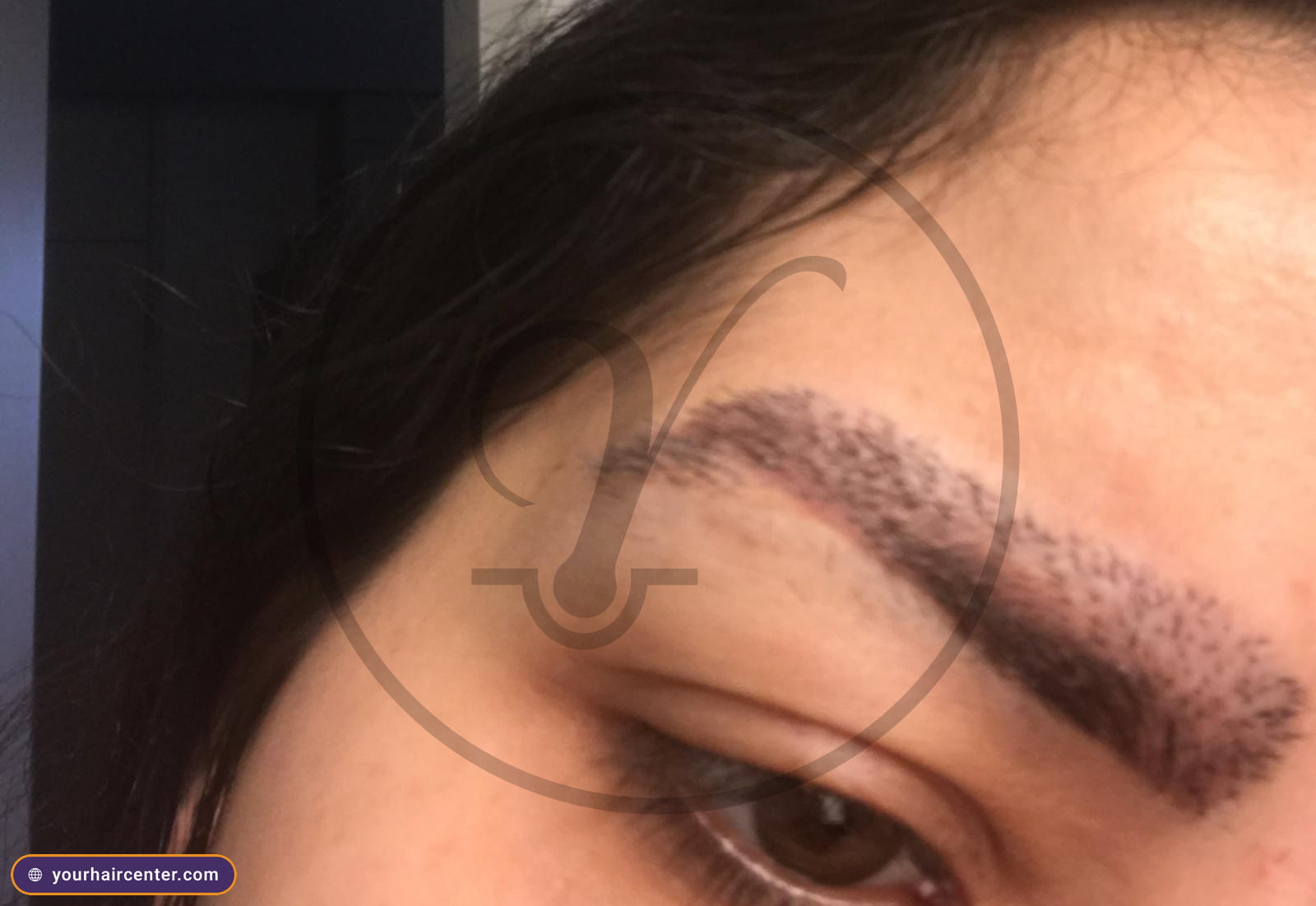
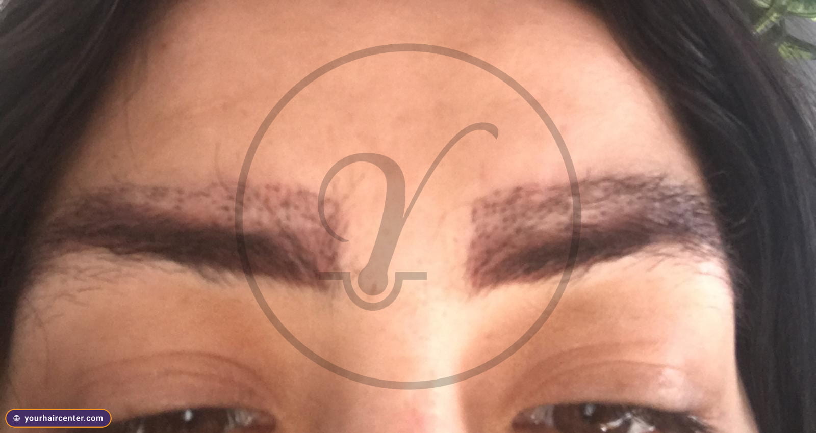
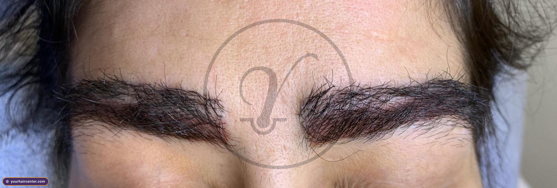
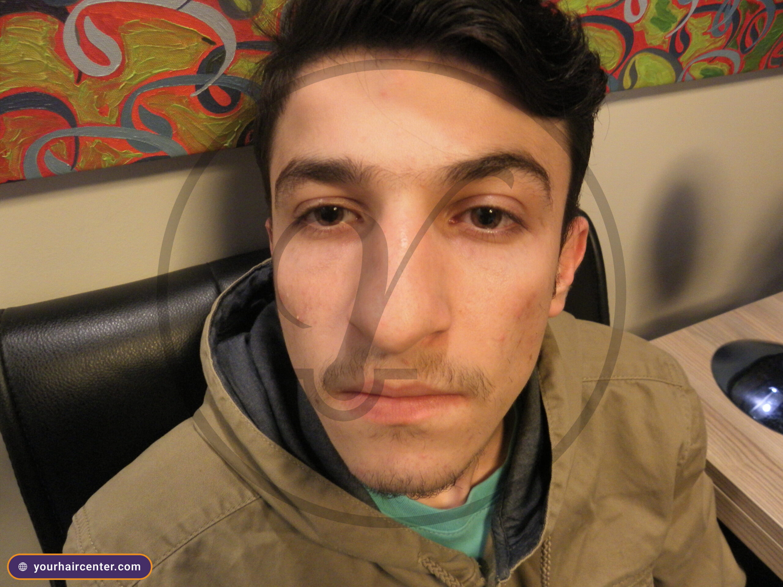
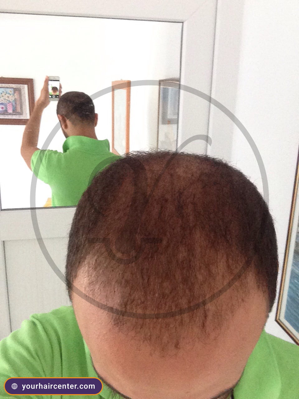
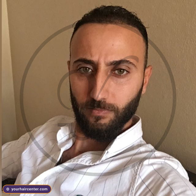
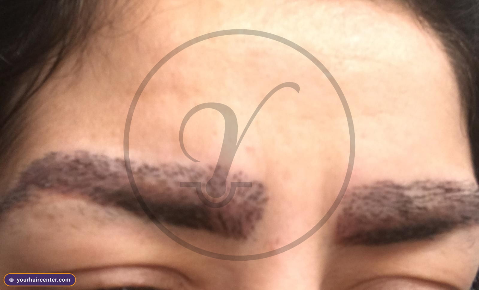
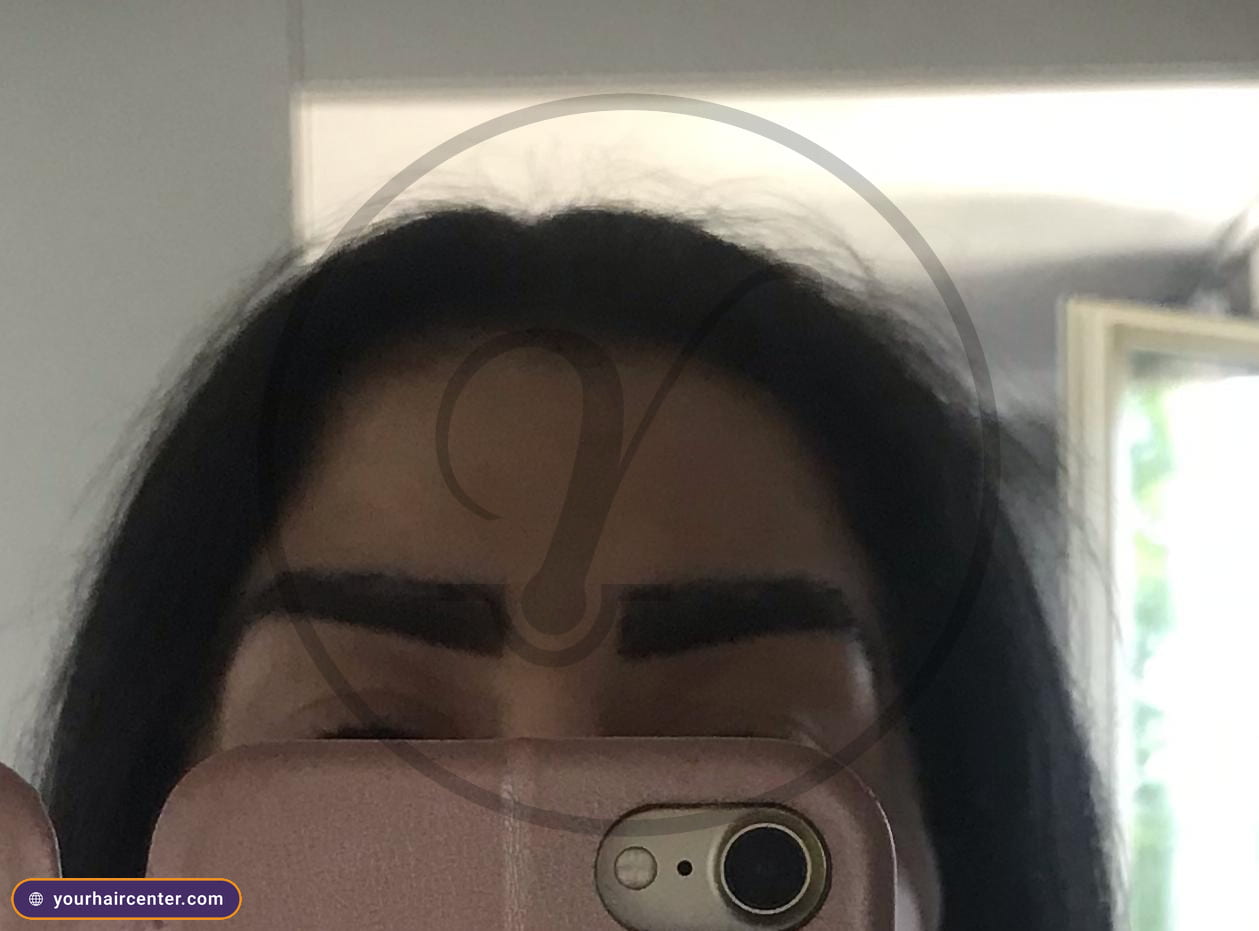
Real results from our patients’ archive. Photos exactly as captured at our accredited partner clinics — no filters, no retouching. Browse more cases in our photo gallery.

Scenario 3, post-partum shedding, the postpartum panel
The picture is a new mother three to six months after delivery, seeing handfuls in the shower, a thinning hairline at the temples, and fine baby hairs regrowing along the front edge as the older shed cohort exits. This is the most common diffuse-shedding scenario in women of reproductive age, and it deserves its own blood test for hair loss rather than the generic one. It is also the heart of what people mean when they search for lab tests for hair loss in females.
This blood test for hair loss panel adds the markers that delivery and lactation put under strain.
- TSH with a reflex free T4 and anti-TPO antibodies, because postpartum thyroiditis affects roughly five to ten percent of postpartum women and is widely missed when the TSH alone reads normal in the early phase.[6]
- Ferritin, since delivery blood loss and the iron demand of lactation routinely drop ferritin below the hair-supporting threshold.[1:1]
- Prolactin, where a level that stays elevated well past the lactation phase points toward a separate workup.
- Twenty-five-hydroxyvitamin D, because prenatal supplementation often stops at delivery while the body’s demand continues.[3:1]
A note for the partner doing the research. Postpartum shedding usually peaks around month four, and for most women fullness returns by around the child’s first birthday, once the trigger labs come back clean and the follicles cycle back into active growth.[7] If month twelve arrives and the part line is still widening, the same blood test for hair loss results re-route the case into the AGA workup above. The hair and scalp analysis gives that trichoscopy-plus-bloods baseline so the trajectory is documented rather than guessed.
Scenario 4, patchy round spots, the autoimmune workup
The picture changes completely here. Instead of even thinning, there are one or more smooth, round, coin-sized bald patches with no obvious scaling, and the patch edges often show short “exclamation-point” hairs on close inspection. This is the alopecia areata presentation, and here the blood test for hair loss logic is autoimmune rather than nutritional. It is the situation people have in mind when they ask what autoimmune labs for hair loss are worth running.
This panel leans on antibody and inflammation markers rather than nutrient stores.
- Anti-thyroid peroxidase (anti-TPO) and anti-thyroglobulin (anti-Tg) antibodies, because autoimmune thyroid disease is the single most common autoimmune condition seen alongside alopecia areata, found in about 7 percent of patients in one large series, with autoimmune conditions of some kind in roughly 12 percent.[8]
- Antinuclear antibody (ANA), which screens for systemic autoimmune overlap such as lupus or scleroderma.
- C-reactive protein (CRP) and erythrocyte sedimentation rate (ESR), inflammation markers that help contextualize an active immune flare.
- A full thyroid panel, TSH with free T4 and free T3, since autoimmune thyroid pathology is the most common comorbidity.[8:1]
- Vitamin D, worth checking as part of the autoimmune workup, since deficiency is common and readily corrected.[3:2]
The next step is explicit. Alopecia areata management belongs to a dermatology team, and the hair loss treatment service routes the patient into in-clinic options once this blood test for hair loss and the trichoscopy localize the picture. Alopecia areata is not a transplant condition during an active flare, so the order of operations matters.
Scenario 5, thinning plus unexplained symptoms, the broad workup
The picture is diffuse shedding plus at least one off-target symptom, persistent fatigue, weight change, mood change, a shift in your cycle, joint aches, brain fog, or gut symptoms. When the shedding is one signal among several, it is the tip of an iceberg and the panel has to widen accordingly.
This blood test for hair loss panel widens well beyond the core five to chase the systemic signal.
- ESR with CRP, for a broad read on inflammation.
- Comprehensive metabolic panel (CMP), covering kidney, liver, electrolytes, and glucose.
- Full iron studies, ferritin with serum iron and total iron-binding capacity (TIBC), rather than ferritin alone.[1:2]
- Vitamin B12 with folate, both of which are independently flagged in shedding.[9]
- Reverse T3 with a full thyroid panel, to capture the conversion-stress thyroid picture a standard TSH can miss.
- Zinc and selenium, the two trace minerals most often investigated alongside iron, though the evidence for testing them routinely is mixed.[9:1]
- HbA1c with fasting insulin, a metabolic-syndrome screen, especially useful in women presenting with crown thinning alongside hirsutism.
The framing for the patient, when shedding is one of several symptoms the panel is wider and the interpretation belongs to internal medicine and a hair specialist working together. The framing for the partner, this is the blood test for hair loss to push for when someone has been told “your bloodwork is normal” but the symptoms have not actually resolved. The hair and scalp analysis is the in-clinic destination that pairs that wider panel with trichoscopy so nothing falls through the gap between specialties. Once specific deficiencies are mapped, hair mesotherapy can deliver the missing nutrients directly to the follicle roots as part of the plan.
Male versus female, what is actually different
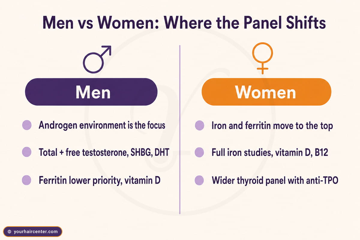
The panel is genuinely gender-skewed, and naming that openly is the part most generic guides skip. The table below shows where the emphasis flips between a typical blood test for hair loss in men and the same workup in women.
| Panel emphasis | Men | Women |
|---|---|---|
| Androgen environment | Total and free testosterone, SHBG, DHT where available, with dihydrotestosterone read as the dominant driver of miniaturization | Total and free testosterone, free-androgen index, DHEA-S, prolactin, plus 17-OHP if PCOS or late-onset CAH is suspected |
| Iron and nutrient stores | Ferritin (often deprioritized but real in vegans and endurance athletes), B12 if vegan, vitamin D | Ferritin (high priority given menstrual loss and reproductive demand), full iron studies, vitamin D, B12 |
| Thyroid and reproductive hormones | TSH with free T4 if symptomatic, prolactin only with galactorrhea | Full thyroid panel with anti-TPO, prolactin, estradiol if the cycle has changed, postpartum panel where applicable |
In men the androgen read is the center of gravity, since dihydrotestosterone is the main pattern-loss driver. In women iron and ferritin move to the top because menstrual loss and pregnancy quietly drain stores. And the thyroid workup widens for women because autoimmune thyroid disease is more common and more often tied to shedding. The Your Hair Center hair and scalp analysis builds the sex-appropriate blood test for hair loss from the same core five plus add-ons rather than running an identical list for everyone.
Frequently asked questions
What are the big 3 for thinning hair?
Most workups lead with three tests, ferritin, thyroid-stimulating hormone, and twenty-five-hydroxyvitamin D, and these three are the backbone of any blood test for hair loss. Ferritin captures your iron stores, TSH captures thyroid drive, and vitamin D captures the most common nutrient deficiency tied to shedding. Together they explain the majority of reversible diffuse-shedding cases, and an abnormal result on any one of them shapes the next step.
What hormone am I lacking if my hair is falling out?
There is no single hormone behind every case. The most common candidates are thyroid hormone (a low free T4 with a normal-range TSH is the classic miss), estrogen (postpartum and perimenopausal drops trigger shedding), and on the surplus side androgens such as DHT and free testosterone, which drive patterned miniaturization. A complete blood test for hair loss should cover thyroid, sex hormones, and androgens together rather than chasing one suspected hormone.
What autoimmune labs for hair loss?
The core autoimmune workup is anti-thyroid peroxidase antibodies (anti-TPO), antinuclear antibody (ANA), and a full thyroid panel of TSH with free T4 and free T3. C-reactive protein and erythrocyte sedimentation rate add an inflammation read. This is the autoimmune arm of a blood test for hair loss, and it matters most when the shedding shows up as patchy round spots or alongside a known autoimmune diagnosis.
How do I tell if my DHT levels are high?
DHT itself is not always offered as a lab, so a blood test for hair loss usually reads it indirectly. Most clinicians measure total and free testosterone with sex hormone-binding globulin (SHBG) and infer DHT activity from the androgen environment plus the clinical picture, with trichoscopy showing miniaturization at the temples and crown. A direct serum DHT assay exists at some labs, with the reference range varying by methodology.
How much does a blood test for hair loss cost?
Costs vary widely by region, lab, and panel breadth. A focused core panel is typically modest out-of-pocket, while broader scenario-specific panels cost more. In the US, insurance coverage depends on the diagnostic code, a hair-loss-only order is commonly classified as cosmetic and denied, while an order tied to a thyroid, anemia, or hormone diagnosis usually clears. An HSA or FSA can often cover the out-of-pocket portion when the order carries a qualifying diagnosis.
Do children need a blood test for hair loss?
Yes, when the presentation is atypical for the child’s age, patchy areata, diffuse loss, or trichotillomania concerns. A pediatric blood test for hair loss still anchors on ferritin, TSH, and vitamin D, with autoimmune add-ons such as anti-TPO and ANA reserved for patchy presentations. The interpretation belongs to a pediatric dermatologist rather than a hair-restoration clinic.
The takeaway and your next step
The five core tests anchor every workup, and the scenario you are living, sudden diffuse, gradual patterned, postpartum, patchy, or thinning with other symptoms, decides which add-ons actually matter. Choosing the right blood test for hair loss is about matching the panel to the pattern, then reading those results next to what the follicles are doing. Your Hair Center runs the full blood test for hair loss paired with digital trichoscopy in a single visit, with a same-day report and a treatment plan matched to the findings. Start with the hair and scalp analysis page, or message the team directly for a free evaluation with the full blood panel.
References
- Diagnostic Value of Serum Ferritin for Telogen Effluvium, a cross-sectional comparative study. PMC. https://pmc.ncbi.nlm.nih.gov/articles/PMC7882421/ (opens in new tab) ︎ ︎ ︎
- Central Hypothyroidism. PMC. https://pmc.ncbi.nlm.nih.gov/articles/PMC3114669/ (opens in new tab) ︎
- Vitamin D Deficiency. StatPearls, NCBI Bookshelf. https://www.ncbi.nlm.nih.gov/books/NBK532266/ (opens in new tab) ︎ ︎ ︎
- Telogen Effluvium. StatPearls, NCBI Bookshelf. https://www.ncbi.nlm.nih.gov/books/NBK430848/ (opens in new tab) ︎
- Trichoscopy of Androgenetic Alopecia, a Systematic Review. PMC. https://pmc.ncbi.nlm.nih.gov/articles/PMC11012765/ (opens in new tab) ︎
- Postpartum Thyroiditis, Causes, Symptoms and Treatment. Cleveland Clinic. https://my.clevelandclinic.org/health/diseases/15294-postpartum-thyroiditis (opens in new tab) ︎
- Hair Loss in New Moms. American Academy of Dermatology. https://www.aad.org/public/diseases/hair-loss/insider/new-moms (opens in new tab) ︎
- Prevalence and Risk Factors Associated with the Occurrence of Autoimmune Diseases in Patients with Alopecia Areata. PMC. https://pmc.ncbi.nlm.nih.gov/articles/PMC8473714/ (opens in new tab) ︎ ︎
- A comprehensive investigation of biochemical status in patients with telogen effluvium. PMC. https://pmc.ncbi.nlm.nih.gov/articles/PMC11626366/ (opens in new tab) ︎ ︎